
JPK Instruments AG, Nanotechnology for Life Science, Bouchéstr. 12, D-12435 Berlin, Germany
e-mail: loebbe@jpk.com
URL: http://www.jpk.com
|
Christian Löbbe
JPK Instruments AG, Nanotechnology for Life Science, Bouchéstr. 12, D-12435 Berlin, Germany e-mail: loebbe@jpk.com URL: http://www.jpk.com |
|
During the last decade biomedical and pharmaceutical applications became more and more common in scanning probe microscopy. Easy sample preparation that requires no freezing or coating of samples made biological samples easy to be imaged in their natural environment. Today atomic force microscopy (AFM) is a well established technique in biology and physiology. Apart from soft matter high resolution imaging, also mechanical properties of living cells can be studied.
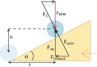
In a cell motility study the AFM cantilever was used as a force sensor for cell (keratocyte) movement. Depending on the substrate (collagen) we cultivated cells in our AFM fluid cell and used the cantilever as a force sensor. The tipless cantilever was equipped with a 2 µm polystyrene bead and was rested on the substrate waiting for a keratocyte crawling under it. The resulting cantilever deflection was measured and the typical pressure applied by the cell’s lamellipodium is calculated. From force distance curves elasticity moduli were calculated. From the slope of the trace curves we got elasticity data, as well as the pushing force of the cell (data courtesy of Josef A. Käs, Soft Matter Physics, University of Leipzig).
![]()