Use of Sub-Micron Metal Rod Decorated Surfaces to Study Basic Cell-Cell and Cell-Surface Interactions


Ulrike Gimsa1, Veronika Kralj-Iglic2, Aleš Iglic3, Stefan Fiedler4, Michael Zwanzig4, and Jan Gimsa5

1Medizinische Fakultät der Universität Rostock, Klinik und Poliklinik für Neurologie, D-18147 Rostock, Germany,
2Institute of Biophysics, Faculty of Medicine, University of Ljubljana, Slovenia
3Laboratory of Applied Physics, Faculty of Electrical Engineering, University of Ljubljana, Slovenia
4Fraunhofer IZM Berlin, Dept. Board Interconnection Technologies, D-13355 Berlin, Germany, and
5Institut für Biowissenschaften der Universität Rostock, Lehrstuhl für Biophysik, D-18059 Rostock, Germany

e-mail: ulrike.gimsa@med.uni-rostock.de

URL: http://neurobiology.med.uni-rostock.de/junior_research_group/index.html
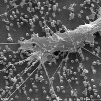
 

We have used sub-micron metal rod decorated surfaces, “nano-lawn” structures, as a substrate to study cell-cell and cell-surface interactions of primary murine astrocytes. Astrocytes are the major cell group of the brain comprising about 50 % of the cells. They support neurons both physically as a cell matrix and physiologically by providing a stable microenvironment and growth factors. Astrocytes form multicellular syncytia in vivo that ensure neuronal homeostasis by taking up excess neurotransmitters and buffering the ionic content of the extracellular medium in the brain. Using sub-micron wires as the matrix for differentiation, we could observe how astrocytes form nanotubular protrusions to make contact with the matrix and each other. The thin tubular structures were very similar to those previously described in pure phospholipid systems and in erythrocytes. Furthermore, cellular material travelling via these nanotubes in transport gondolas has been observed. It could be theoretically shown that curvature-induced self-assembly of interacting anisotropic membrane components may lead to the spontaneous formation of thin nano-tubular membrane protrusions. This self-assembly may represent a relevant physical mechanism of nano-tube formation even if membrane skeleton elements, such as actin fibers, were essential for triggering nano-tube formation in cells.