
Maurice E. Müller Institute, Biocentre, University of Basel, Klingelbergst. 70, CH-4056 Basel, Switzerland
e-mail: Andreas.Engel@unibas.ch
|
Andreas Engel
Maurice E. Müller Institute, Biocentre, University of Basel, Klingelbergst. 70, CH-4056 Basel, Switzerland e-mail: Andreas.Engel@unibas.ch |
|
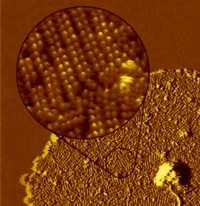
Membrane proteins are embedded in the lipid bilayer and are the doors of the cells. With characteristic dimensions of 5-10 nm they are nanomachines that fulfill key functions such as energy conversion, solute transport, secretion and signal transduction. Their central role in a wide range of diseases may explain the fact that 70% of all drug targets are membrane proteins. The atomic force microscope (AFM) produces images with an outstanding signal-to-noise ratio and addresses single molecules under native conditions, keeping the sample in buffer solution. Progress in sample preparation and instrumentation has led to topographs that reveal sub-nanometer details and surface structure of biomolecules. The AFM can therefore be used to study surface structure and dynamics of membrane proteins in native membranes. Alternatively, two-dimensional crystals of purified membrane proteins are assessed by electron crystallography to elucidate their structure at atomic resolution. Examples presented concern the prototype G-protein coupled receptor rhodopsin and members of the medically important aquaporin family.
![]()